ARRS Case of the Week
CARDIOVASCULAR IMAGING: Pulmonary Artery
Case Author: Travis S. Henry, MD, Emory University School of Medicine
History
66-year-old woman with worsening dyspnea.
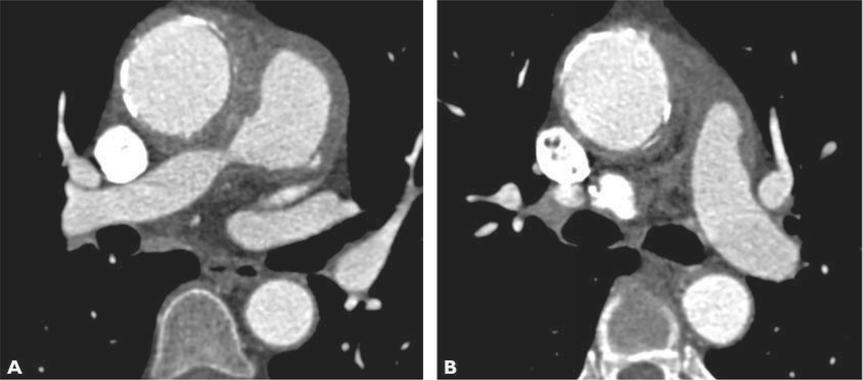
Imaging Findings
Axial CT images (A and B) show circumferential soft tissue around the ascending aorta and central pulmonary arteries. The right main pulmonary artery is severely narrowed (A). Calcification is present within the wall of the aorta and pulmonary artery involving both the intima and media.
This page is updated with new content weekly. It was last updated on February 22, 2021.
You May Also Be Interested In