ARRS Case of the Week
GENITOURINARY IMAGING: Kidney
Case Author: Courtney Coursey Moreno, MD, Emory University School of Medicine
History
44-year-old woman with pain.
Imaging Findings
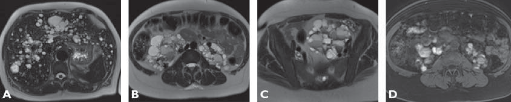
T2-weighted MR image without fat saturation (A) shows numerous rounded primarily hyperintense structures within the liver. T2-weighted image without fat saturation (B) shows numerous rounded primarily hyperintense structures in the right and left renal fossae. Some of the structures also have intermediate and low signal intensity. T2-weighted image without fat saturation (C) shows a left pelvic mass composed of multiple primarily hyperintense rounded structures and intermediate- and low-signal-intensity structures. Contrast-enhanced T1-weighted fat-saturated MR image (D) through the level of the renal fossae shows corresponding rounded areas of low and high signal intensity.
This page is updated with new content weekly. It was last updated on September 16, 2019.
You May Also Be Interested In
|
|
|
Earn 10 CME/SA-CME Credits
Focus on honing your diagnostic skills with this distinctive Online Course that combines didactic lectures and rapid-fire case review sessions to provide deeper insights into abdominal imaging.
Learn more and order.
|