ARRS Case of the Week
ULTRASOUND: Obstetrics
Case Author: Manjiri Dighe, MD, University of Washington Medical Center
History
30-year-old pregnant woman undergoing routine anatomic scan.
Imaging Findings
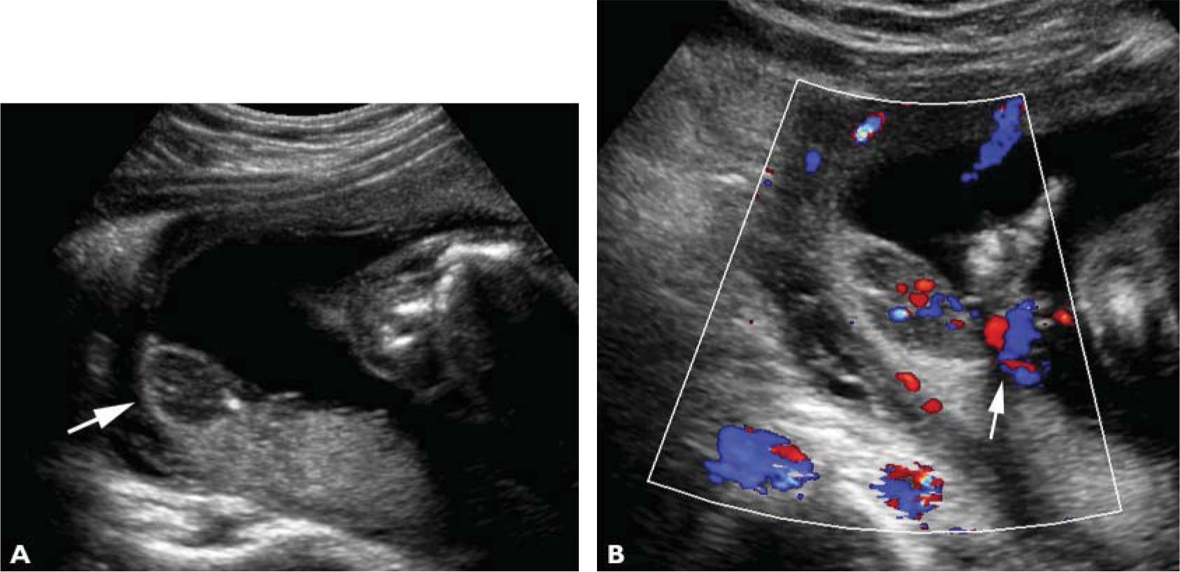
Sagittal B-mode ultrasound image of the placenta (A) shows a hypoechoic mass close to the fetal surface of the placenta (arrow). Sagittal color Doppler ultrasound image of the placenta (B) shows internal vascularity in the mass, which is located close to the cord origin (arrow).
What is the correct diagnosis?
- Chorioangioma
- Fibroid
- Hematoma
- Venous lake
Correct Answer
Chorioangioma
Teaching Points
Chorioangioma is seen as a hypoechoic mass with internal vascularity in the placenta. It is commonly located close to the fetal surface of the placenta near the cord origin. Complications seen with placental chorioangiomas include polyhydramnios, hydrops from arteriovenous shunting, and fetal anemia from hemolysis and intrauterine growth restriction.
The presence of vascularity within the mass differentiates a chorioangioma from a hematoma. Very slow flow can be seen within a venous lake at B-mode imaging, differentiating it from a chorioangioma with arterial flow.
Fibroids are more commonly seen along the maternal side of the placenta, whereas chorioangiomas are more common along the fetal surface of the placenta and close to the cord origin.
Suggested Readings
Bromley B, Benacerraf BR. Solid masses on the fetal surface of the placenta: differential diagnosis and clinical outcome. J Ultrasound Med 1994; 13:883–886
Sepulveda W, Alcalde JL, Schnapp C, Bravo M. Perinatal outcome after prenatal diagnosis of placental chorioangioma. Obstet Gynecol 2003; 102(5 pt 1):1028–1033
This page is updated with new content weekly. It was last updated on August 19, 2019.
You May Also Be Interested In